Porcine ear necrosis in intensive production systems
To better understand porcine ear necrosis and to optimise control measures, it is important to have more detailed information on the prevalence, lesion severity, and risk factors of porcine ear necrosis.
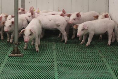
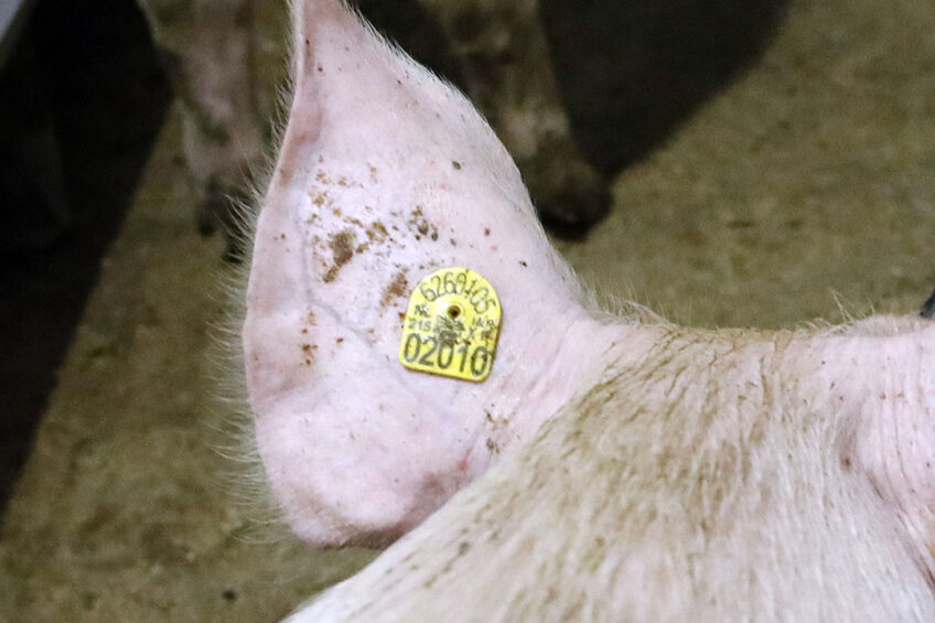
Porcine ear necrosis, also known as ear tip necrosis or ear necrosis syndrome, is described as blue to black discoloration of the ear tip or margin followed by the formation of necrotising ulcerative lesions. Porcine ear necrosis is more prevalent in nursery pigs. Itis an increasing problem in countries with intensive pig production systems.
Potential risk factors of porcine ear necrosis
Potential risk factors of lesion formation include environmental and management conditions. Loss of a part of the ear or the entire ear is possible after healing. However, the precise etiology and pathogenesis of porcine ear necrosis remains unexplored.
Prevalence
Porcine ear necrosis prevalence varies between countries, between farms, and over time within a farm. It ranges from 30 to 70%. Visible ear lesions appear mostly in weaned piglets from 6 to 8 weeks of age. These remain visible until 14-16 weeks of age. Porcine ear necrosis is a multifactorial disorder in which infectious and non-infectious factors may play an important role. Infectious risk factors include viral and bacterial infections. Viruses potentially involved include porcine circovirus type 2 and porcine reproductive and respiratory syndrome virus.
Staphylococcus hyicus is the most frequently isolated bacterium from porcine ear necrosis lesions. In addition, Staphylococcus aureus, Spirochetes of the genus Treponema, and Mycoplasma suis are associated with porcine ear necrosis.
Non-infectious risk factors involved in porcine ear necrosis include fully slatted floor with no straw, poor air quality, high humidity, high stocking density, early weaning, low availability of drinkers and feeders per pig, mycotoxin contamination of the feed, fighting and ear biting, and genetics.
Lesion severity scoring
Diagnosis is based on the presence of lesions on the affected ears. Lesion severity is scored according to the affected surface of the ear based on a method developed by Pejsak and colleagues (2011) from the National Veterinary Research Institute in Pulawy, Poland. According to this scoring method weak changes cover less than 5% of the ear surface, mild lesions cover 5-10% of the ear, and serious lesions cover more than 10% of the ear. In addition, in 2020, Malik and colleagues from Ghent University, Belgium developed a 4-point lesion severity scoring system in which score 1 shows a small crust on ear tip, score 2 is for a small wound on ear tip with reddening around, score 3 means bloody, necrotic wound on ear edge, and score 4 displays partial lack of auricle with necrotic edge.
Pathogenesis and clinical signs
The precise etiology and pathogenesis of porcine ear necrosis remain unknown. 3 hypotheses however have been suggested for disease development. According to first hypothesis, the necrosis is caused by toxins of Staphylococci and starts on the outer surface of the injured skin. The second hypothesis states that ear tip necrosis is due to damage of small blood vessels caused by cold agglutinins or immune complexes produced during infection with Mycoplasma suis. The third hypothesis declares that external trauma such as ear biting or environmental factors cause ear tip necrosis. Necrotic lesions vary from mild to severe. Mild lesions consist of an encrusted sore, localised on the ear tip or ventral margin of the ear which can heal without treatment, and sever lesions include epidermal ulceration and necrotic, moist lesions of the pinna.
Treatment and prevention of porcine ear necrosis
Severely affected pigs need to be housed in a hospital pen to avoid further biting by other pigs. Antibiotic therapy for 2 weeks with amoxicillin, amoxicillin with clavulanic acid, or sulfadiazine-trimethoprim can slow lesion progression. Vaccination of piglets against porcine circovirus type 2 and porcine reproductive and respiratory syndrome virus, adequate pen design, avoiding overcrowding, limiting mixing of pigs, optimal ventilation and air quality, proper feed quality and sanitation reduce porcine ear necrosis prevalence.
Conclusions
Porcine ear necrosis is a common disorder especially in nursery pigs worldwide. However, its exact etiology and pathogenesis remain unknown which hinders optimal treatment. Thus, control and prevention measures should focus on reducing potential risk factors. In addition, further research is required to investigate etiology and pathogenesis of the disorder to elucidate causative factors and to decrease the risk of occurrence.
Join 18,000+ subscribers
Subscribe to our newsletter to stay updated about all the need-to-know content in the pigsector, three times a week.